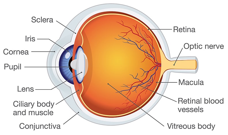
Anatomy of the Eye
The eye is a truly remarkable and complex organ. It is the primary means through which most of us come to recognize and comprehend the world around us. It is a multipurpose instrument that receives light, controls how much light enters the eye, focuses the light, and then sends signals to the brain, which translates the signals into recognizable images. But without light — there is no vision. The eye measures light; whether emitted directly from a source (like a light bulb) or reflected from the surfaces of objects.
The eye has many different parts that work together to perform the various functions of the eye. Some of the parts, described below, are the sclera, cornea, pupil, iris, lens, vitreous humor, retina and optic nerve.


Sclera
The white of the eye. The sclera gives the eye structural strength and protection for the inner elements of the eye.
Cornea
This is the outer clear covering over the center of the eye. The cornea acts like both a window and a lens. Light first enters the eye through the cornea. The shape of the cornea then acts as a lens to begin to bend or “refract” light toward a focal point at the back of the eye. The cornea is the primary focusing element of the eye. It is made up of five layers of material. The inner layers add strength and substance to the eye, while the outermost layer provides a tough, protective membrane that helps to shield the more delicate areas of the eye beneath. This outer layer of the cornea is called “epithelium”. The epithelium is remarkably resilient. Its cells can regenerate in as little as three days. This allows any superficial injury to the outer cornea to heal very quickly.
Pupil
The visible black center of the eye is the pupil. The pupil works like the aperture or opening of a camera to regulate the amount of light entering the eye. In bright light, the pupil closes down to limit how much light comes into the eye. In dim light the pupil opens up and becomes visibly larger. This allows the eye to receive more light.
Iris
From blue to green to brown, and in various shades, the iris gives color to the outward appearance of the eye. But the iris is more than cosmetic; its function is to work with the pupil as the means by which the pupil is opened and closed. The muscles of the iris expand or contract around the pupil, thus changing the size of the opening from larger to smaller.
Lens
This is the clear element just behind the pupil. Because it is flexible, it can change shape from thinner to thicker. This works to change the focal length and thus the focal point of the light passing through it. The lens is actually a kind of secondary focusing component, something like the second lens of a telescope, which fine–tunes the light entering from the cornea so that it focuses precisely on the retina. Due to the aging process, the lens begins to lose its elasticity and its ability to sharply focus light. This is the condition known as “presbyopia” (old eye) which often occurs by age 40 or 50. If the lens becomes clouded and hardened, light not only becomes unfocused, it becomes blocked to a greater or lesser degree. This is the condition described by the term, “cataracts”.
Vitreous Humor
The vitreous body of the eye is a clear, thick, semi–solid material like a gel. It is the inner substance, which gives shape and firmness to the eye. Within this vitreous material small lumps or hairlike pieces known as “floaters” may develop. These bits are so named because they appear to “float” before, on or in front of the eye.
Retina
This is the focusing screen of the eye. The retina is made up of nerve tissue. This keenly sensitive nerve tissue lines the rear wall of the eye. The retina receives the finely focused light from the lens and turns the light energy into electrical impulses. These impulses can then be translated into images by the brain. In a properly functioning eye, light is focused at a point precisely on the retina.
Optic Nerve
The optic nerve is the conductor that transmits the electrical impulses from the retina to the brain. This is the final stage of the process we know as vision. The optic nerve enables the light received by the eye to make sense.
